
Figure 2.
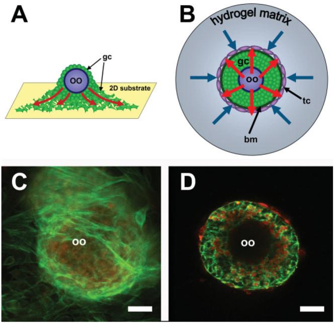
(A) Follicle architecture in a two-dimensional culture system. On a two-dimensional substrate, proliferating follicle cells spread onto the surface (red arrows). (B) In a three-dimensional culture system, the follicle grows radially from the oocyte (red arrows). If a matrix surrounds the follicle, it will produce an opposing force on the follicle (blue arrows). This is demonstrated in (C) and (D). (C) Follicles cultured on a two-dimensional substrate lose their structure as they spread onto the surface, whereas (D) follicles maintain their morphology in an alginate matrix. Follicles in (C) and (D) are labeled with dextran (red) to demonstrate active uptake of compounds from the culture media and phalloidin (green) to visualize the distribution of the actin cytoskeleton of follicular cells. oo, oocyte; gc, granulosa cells; tc, theca cells. Scale bar =30 μm. (Figures 2C and 2D are reprinted from Kreeger, PK, Deck, JW, Woodruff, TK, Shea, LD. The in vitro regulation of ovarian follicle development using alginate-extracellular matrix gels. Biomaterials 2006;27:714–723, with permission from Elsevier.)