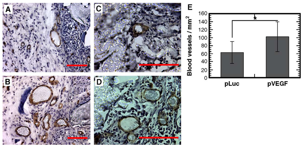
FIG. 7.
Immunohistochemical staining with CD31 (PECAM-1) monoclonal antibody to identify blood vessels. Sections were counterstained with hematoxylin. Scaffolds releasing (A, C) pLuc or (B, D) pVEGF. Images captured immediately adjacent to the scaffold (A, B) or within the scaffold (C, D). Original magnification for photomicrographs: (A, B) 200×, (C, D) 400×. Scale bar, 100 µm. (E) Blood vessel density within tissue sections containing the polymer scaffold (n = 3). *Statistical significance at P < 0.001.

