Figure 3.
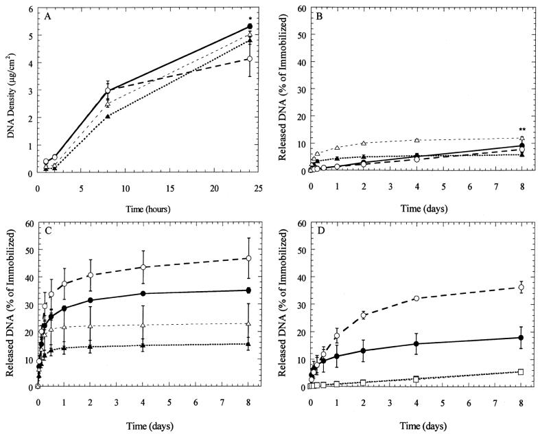
DNA deposition and stability. (A) Polyplexes (circles) and lipoplexes (triangles) were deposited onto polystyrene (●, ▲) and serum-coated polystyrene (○, △) for 24 h. Substrates were incubated with 2 μg of DNA; (B) immobilized polyplexes and lipoplexes were exposed to phosphate-buffered saline (PBS, pH 7.4); (C) conditioned media; (D) polyplexes incubated with cell growth media (●, PS; ○, FBS-PS), and trypsin (■, PS; □, FBS-PS) at 37°C. Data are presented as average ± standard deviation of the mean. *P < 0.05 relative to FBS-PS deposition of polyplexes after 24 h; **P × 0.05 relative to lipoplexes released from PS at 8 days.

