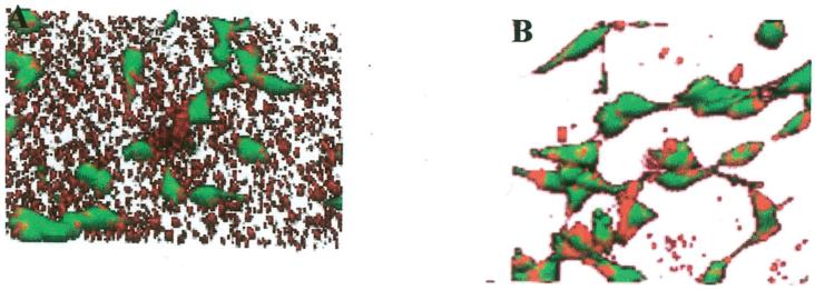
Figure 8.
Distribution of cells and polyplexes on substrate following cell culture. Confocal microscopic images of polyplexes on (A) PS and (B) FBS-PS 20 h after cell seeding. Polyplexes (2 μg, N/P = 25) were incubated on substrates for 2 h followed by washing and cell seeding. Images were captured 20 h after cell seeding. Plasmid was labeled with tetramethylrhodamine, and cells were stained with fluorescein diacetate.

