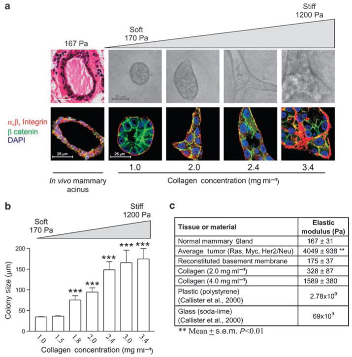
Figure 1.
MEC growth and morphogenesis reflect changes in matrix stiffness. (a) Confocal images of MEC grown in 3D cultures. As MECs grow in progressively stiffened matrices (170–1200 Pa), MEC morphology becomes progressively disrupted. Irregular MEC changes are characterized by disrupted cell–cell adherens junctions and tissue polarity, illustrated by a loss of β-catenin (green) and loss of β4 integrin (red) organization (nuclei =blue). (b) Normal MEC acini reaches a proliferative growth-arrested phase when cultured in soft gels that is lost as they are cultured in stiffer matrices. (c) Measured elastic modulus for a variety of substrates. Values represent the mean ± s.e.m. of four measurements from multiple mice and gels. **P≤0.01, ***P≤0.001. (Reproduced with modification and proper permission obtained from Elsevier as published in Paszek et al., 2005.)

