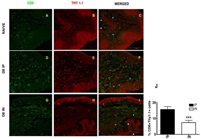
Figure 2.
Confocal microscopy confirms differences in the magnitude of the Ag-specific CD8 T cell response in the vagina on day 8. Frozen sections of the vaginal tract were fixed and stained with CD8 and Thy 1.1 Abs. Thy1.1 staining shows Ag-specific CD8 T cell staining in the stroma of the vaginal mucosa on day 8 postinfection with LCMV. A, D, and G, CD8 T cells in green and B, E, H, Thy1.1 staining in red. C, F, I, Merged image shows Thy1.1 staining only on CD8 T cells of infected mice. Naive stained only for CD8 T cells. Images were captured using a 20× objective lens. The epithelial layer is indicated by the white arrowheads (luminal edge) and blue arrowheads (basement membrane). J, Percentage of Ag-specific CD8 T cells in vaginal tract of infected mice at day 8 postinfection. Thy1.1+ CD8+ cells were counted on blinded samples.***, p < 0.001.

