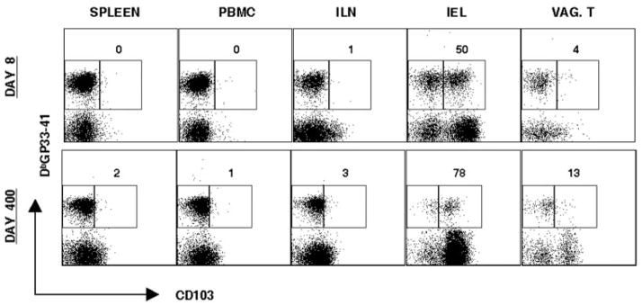
Figure 5.
Expression of CD103 on Ag-specific CD8 T cells in genital mucosa. Lymphocytes were isolated from the spleen, PBMC, ILN, IEL, and vaginal tract from day 8 and day 400 LCMV Armstrong i.p. Cells were stained with CD8, CD103, and DbGP33–41. Representative staining is shown, gating on CD8 T cells. Numbers represent percentage of CD103high Ag-specific CD8 T cells in indicated tissue compartments. For vaginal tract, tissues are pooled from four to six mice.

