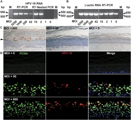
Figure 3.
Infectivity assays of HPV-18 virions in PHKs. HPV virions were titered by quantitative real-time PCR. (A) HPV cDNA detection in submerged PHK cultures after infection at the indicated MOI. A 521-bp cDNA fragment of a spliced early viral mRNA was detected by ethidium bromide staining after RT–PCR (left panel) or RT-nested PCR (right panel). (B) A 642-bp-long β-actin cDNA served as an internal control. (M) 50-bp ladder. (C) Immunohistochemical detection of the HPV major capsid protein L1 (reddish brown) in 14-d raft cultures of PHKs infected with HPV virions.(Left panel) MOI of 800 or higher. (Middle panel) MOI of 50 up to 400. An uninfected culture is shown in the right panel. Arrowheads point to the boundary between the upper cornified strata and live epithelium below. (D) Four-micron sections from the raft cultures above were probed for PCNA (Alexa Fluor 488, green) and for viral DNA (Cy3, red). Cellular DNA was revealed by DAPI (blue).

