Figure 3.
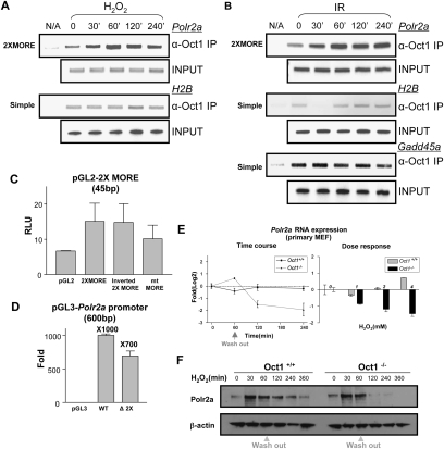
Oct1 functionally regulates Polr2a. (A) Time-course ChIP assay of Polr2a using HeLa cells treated with H2O2. H2B and input DNA are shown as controls. (B) Similar time-course experiment using IR. Oct1 association with a simple octamer site in Gadd45a is additionally shown. (C) Reporter activity of a 2XMORE linked to the SV-40 promoter in a luciferase-based assay. Constructs were transfected into HeLa cells. Experiment was performed in triplicate. Error bars depict standard deviations. (D) Activity of the human Polr2a promoter fragment (−600 to +1 relative to the TSS) linked to luciferase. A 45-bp 2XMORE deletion was made in the context of the full sequence. Empty vector was used as a control. (E, left panel) Real-time RT–PCR using intron-spanning mouse Polr2a primers and either wild-type or Oct1-deficient MEFs. Message levels are shown following treatment with 2 mM H2O2 relative to wild-type fibroblasts under unstressed conditions on a log2 scale. (Right panel) Dose response experiment. Polr2a expression was measured 4 h following exposure to different H2O2 doses. (F) Western blot showing time course of Pol II large subunit expression in immortalized MEFs cells treated with H2O2. β-actin is shown as a loading control.

