Figure 7.
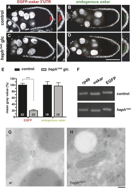
PTB promotes copackaging of oskar mRNA molecules in vivo. In situ hybridization of heterozygous (A,B) and homozygous (C,D) heph1545 late stage 9 egg chambers expressing a UAS-EGFP-osk 3′UTR construct under the control of the mat-α4-tub-Gal4 driver. (A,C) Egg chambers double-stained for EGFP transcripts (red) and DAPI (white). Images A and C were taken using identical settings. (B,D) Egg chambers double-stained for endogenous oskar transcripts (green) and DAPI (white). Endogenous oskar was detected using a probe exclusively covering the coding region. Pictures B and D were taken using identical settings. Bar, 60 μm. (E) Quantification of the in situ hybridization signal detected at the posterior pole of late stage 9 oocytes. Intensity values obtained using ImageJ are expressed as a percentage of control oocytes. (***) P < 0.001 in a Student's t-test. (F) Expression levels of rp49, endogenous oskar, and EGFP-oskar 3′UTR transcripts in both heterozygous and homozygous heph1545 backgrounds, as measured by semiquantitative RT–PCR. (G,H) Electron micrographs of w (G) and heph03429 (H) early stage 9 oocytes hybridized with an oskar antisense probe. oskar mRNA was detected using 10-nm gold particles, and the central region of the oocyte is shown. Note that in heph03429 oocytes, the slightly electron-dense oskar RNP complexes are smaller and more numerous. Bar, 150 nm.

