Figure 6.
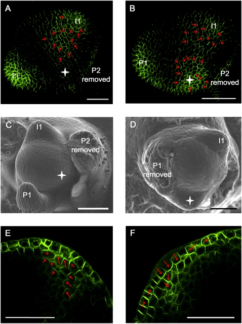
PIN1 reacts to ectopic auxin application. (A,B) Maximal projection of transversal confocal scans of wild-type tomato meristems expressing AtPIN1:GFP. (A) Control meristem 20 h after microapplication of DMSO only, showing a normal expression pattern with AtPIN1:GFP polarizing toward I1. Extending from this position is a PIN1 expression domain with polarity clearly away from I2 and toward I1. (B) Comparable meristem, 20 h after microapplication of IAA in DMSO. A convergence point is visible at I1 as well as at the site of microapplication. (Red arrows) AtPIN1:GFP polarity. (C,D) Scanning electron microscope pictures of the same meristems as shown in panels A and B, respectively, 50 h after treatment. (D) Note ectopic primordium formation at the site of IAA microapplication (white star). (E,F) Optical longitudinal confocal section through IAA-induced PIN1 convergence point, 10 h (E) and 20 h (F) after microapplication. Note the apical (E) followed by basal (F) polarization of AtPIN1:GFP in inner tissues. Bars, 50 μm.

