Figure 4.
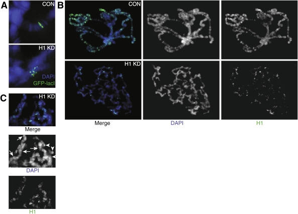
Effects of H1 depletion on alignment of sister chromatids in polytene chromosomes. (A) H1 depletion results in misalignment of chromatin fibrils in an interband region in 61F. The genomic position of a P-element insertion containing 256 lacO sites was visualized by indirect immunofluorescence (IF) through binding of an ectopically expressed GFP-lacI protein to the locus in polytene chromosomes. In control squashes (pINT-1-Nau + Tubulin-GAL4, CON), the GFP signal was observed as a single straight band, indicative of an alignment of endoreplicated chromatin fibrils. In H1-depleted polytene chromosomes (pINT-1-H15M + Tubulin-GAL4, H1 KD), the GFP signal was dispersed into multiple isolated spots, indicating the loss of perfect alignment. The animals were incubated at 29°C throughout their life cycle. (Blue) DAPI; (green) GFP-lacI. (B) Residual H1 protein in H1-depleted polytene chromosomes is unevenly distributed and correlates with regions of persistent polytene band–interband structure. Polytene chromosome squashes from control (pINT-1-Nau + Tubulin-GAL4, CON) and H1-depleted (pINT-1-H15M + Tubulin-GAL4, H1 KD) salivary glands were stained with the H1 antibody. In control chromosomes, H1 localizes primarily to bands in euchromatic arms and to the pericentric region. In H1-depleted larvae, the H1 signal is dramatically reduced, and the residual protein is distributed over a limited number of loci. The majority of these loci also have a partially conserved polytene band–interband structure, as evidenced by DAPI staining. In contrast, chromosome regions that do not contain detectable H1 typically exhibit amorphous, clumped morphology. The animals were incubated at 29°C throughout their life cycle. (Blue) DAPI; (green) H1. (C) In H1 knockdown larvae, polytene band–interband structure is partially preserved in loci with elevated residual H1. Whereas DAPI stains equally brightly the residual structured bands (arrows) and unstructured chromatin clumps (arrowheads), H1 staining predominantly has an appearance of discrete bands. No diffuse signal can be observed for the H1 staining. Furthermore, there is a substantial overlap between the residual DAPI and H1 bands.

