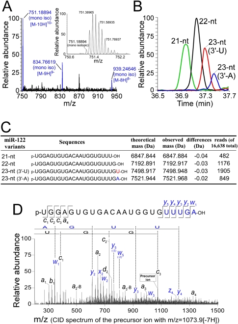
Figure 1.
Mass spectrometric characterization of miR-122 isolated from mouse liver. (A) Mass spectrum of the 23-nt (3′-A) variant of miR-122. The multiply charged negative ions, [M-8H]8−, [M-9H]9−, and [M-10H]10−, of deprotonated miR-122 can be seen. (Inset) A series of isotopic ions of [M-10H]10− are shown in the magnified graph; the monoisotopic ion (m/z 751.19016) is indicated. The mass spectrum was acquired in the range of m/z 750–1500 with mass resolution of 30,000 (FWHM). (B) Mass chromatograms shown by [M-8H]8− and [M-9H]9− of each miR-122 variant, 21-nt (green), 22-nt (black), 23-nt (3′-U), (red) and23-nt (3′-A) (blue). (C) Sequences and exact molecular masses of the miR-122 variants. The read number from the pyrosequencing for each variant is indicated. The total number of reads was 16,638. (D) CID spectrum of the 23-nt (3′-A) variant of miR-122. [M-7H]7− (m/z 1073.9) was selected as a precursor mass for CID. The product ions were assigned according to Mcluckey et al. (1992). Partial sequences from both termini determined by this spectrum are indicated.

