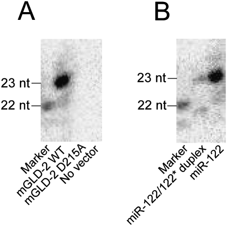
Figure 4.
In vitro 3′ adenylation of miR-122 by immunoprecipitated mGLD-2. (A) The synthetic miR-122 was 3′-adenylated in vitro by the immunoprecipitated mGLD-2 in the presence of [α-32P]ATP. Wild-type mGLD-2 (mGLD-2 WT) or its inactive mutant (mGLD-2 D215A) was immunoprecipitated with anti-Flag M2-agarose beads from the lysate of Huh7 cells transfected with pcDNA3 Flag-HA-mGLD-2 or its inactivated mutant form (pcDNA3 Flag-HA-mGLD-2 D215A). No vector represents a negative control preparation from the same cell lysate untransfected. [α-32P]AMP-labeled miR-122 was analyzed by 20% denaturing PAGE and visualized by an imaging analyzer (BAS5000; FujiFilm). The marker is the 5′-32P-labeled miR-122 (22-nt). (B) The synthetic miR-122 (single strand) and miR-122/mi-R122* duplex were 3′-adenylated in vitro by the immunoprecipitated mGLD-2 in the presence of [α-32P]ATP. The products were analyzed as described in A.

