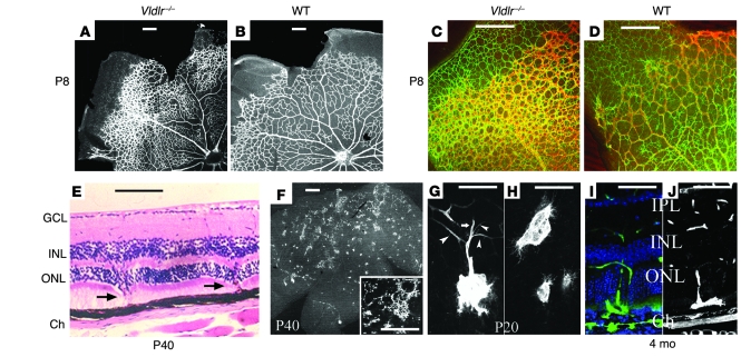
Figure 1. Intraretinal vascular abnormalities in Vldlr–/– mouse retinas.
(A and B) The developing superficial vascular plexus of Vldlr–/– retinas was hyperdense, particularly in the periphery, at P8. (C and D) Peripheral hypervascularity (isolectin GS; red) was associated with increased density of astrocytes (GFAP; green). (E) H&E-stained retinal sections from a P40 Vldlr–/– mouse showed intraretinal vessels (arrows) that originated from the inner retinal vascular plexuses and migrated through the photoreceptors to the subretinal space. GCL, ganglion cell layer; Ch, choroid. (F) These intraretinal vessels formed retinal-retinal anastomoses throughout the central two-thirds of the Vldlr–/– retinas. Inset shows a higher-magnification view. (G) Intraretinal vessels originated from multiple sprouts off the deep (arrowheads) and intermediate vascular plexuses (arrow) and could form large angiomatous structures within the subretinal space. (H) Filopodia extended from subretinal vascular endothelial cells and extended laterally to form retinal vascular anastomoses. (I and J) Fluorescein angiography (I) or isolectin GS staining (J) demonstrated the retinal origin of the intraretinal and subretinal NV. IPL, inner plexiform layer. Scale bars: 100 μm.

