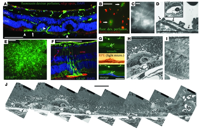
Figure 2. Characterization of abnormal NV in the Vldlr–/– mouse retina.
(A) Retinal cross-section montage demonstrating multiple retinal abnormalities in the Vldlr–/– mouse retina at 3 months of age, including subretinal vessels (arrowheads), vascular leak (arrow), abnormal retinal morphology, abnormal neuronal layering in the ONL and INL, and loss of red/green (rd/gr) cone opsin staining. (B and C) Vascular leak was associated with a small subset of subretinal vessels, as demonstrated by extravasation of perfused 43-kDa fluorescein-labeled dextran. (D) Fenestrae within endothelial cells of the subretinal vessels were observed by electron microscopy. Inset shows a lower-magnification view, in which the boxed region defines the bounds of the higher-magnification view. (E) Punctate GFAP staining in retinal whole mounts demonstrated glial activation associated with abnormal NV in the Vldlr–/– mouse retinas. (F) This punctate staining represented Müller cells specifically activated around the abnormal intraretinal vessels. (G and H) The RPE formed multicellular layers that engulfed abnormal intraretinal vessels. (I) Electron microscopy demonstrated normal retinal morphology at P12, just prior to NV onset. (J) Montage of multiple electron microscopy images showing retinal damage spatially associated with abnormal NV in 2 separate regions, while intermittent retinal morphology between lesions remained mostly normal. Scale bars: 100 μm (A–C and E–G); 50 μm (H–J); 10 μm (D).

