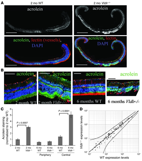
Figure 6. Evidence of oxidative stress associated with subretinal NV in Vldlr–/– mouse retinas.
(A) Acrolein staining increased in Vldlr–/– retinas compared with age-matched WT controls. Acrolein staining in Vldlr–/– retinas was largely within the central retina, where intraretinal NV is most prominent. Panels are composite montages of multiple serial micrographs. (B) Higher-magnification images demonstrating that acrolein staining localized to the photoreceptor layer and INL. (C) Acrolein staining significantly increased in 2-mo-old Vldlr–/– compared with WT retinas. At 6 mo of age, acrolein staining was similar in the peripheral retinas of WT and Vldlr–/– mice (P = 0.245), but was significantly stronger in the central regions of Vldlr–/– retinas, where subretinal NV occurs, compared with retinas of WT mice. Error bars denote SEM. (D) Expression of genes involved in oxidative stress defense mechanisms was similar between P21 Vldlr–/– and WT retinas. Solid line indicates equivalent expression levels; dotted lines represent 95% confidence level ranges. Scale bars: 500 μm (A); 50 μm (B).

