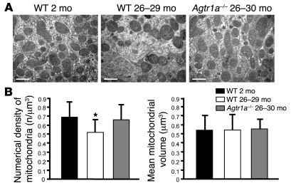
Figure 6. AT1A deficiency prevents loss of mitochondria.
(A) Representative transmission electron micrographs of the ultrastructure of mouse proximal tubular cells obtained from resin-embedded kidney sections from young wild-type mice (2 months old), aged wild-type mice (26 to 29 months old), and Agtr1a–/– animals (26 to 30 months old). Scale bar: 1 μm. (B) The number of mitochondria per volume in proximal tubular cells in aged wild-type animals was decreased with respect to wild-type 2-month-old and Agtr1a–/– mice. Agtr1a–/– animals showed the same numerical density as wild-type 2-month-old animals. *P < 0.01 versus wild-type 2-month-old and Agtr1a–/– animals by ANOVA corrected with Bonferroni coefficient. Mean mitochondria volume evaluated on the same transmission electron micrographs through morphometrical analysis did not differ among groups. Data are mean ± SD.

