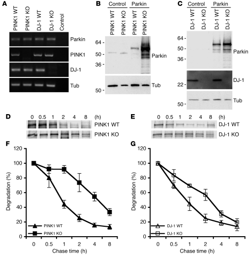
Figure 7. Genetic ablation of mouse Pink1 or Dj-1 results in increased stability of aberrantly expressed Parkin.
(A) Expression of Parkin, PINK1, and DJ-1 in PINK1- or DJ-1–deficient mouse fibroblasts. RT-PCR detection of Parkin, PINK1, and DJ-1 in PINK1 WT, PINK1 KO, DJ-1 WT, and DJ-1 KO cells. Control, no cDNA template added. (B and C) Increased accumulation of aberrantly expressed Parkin in PINK1 KO and DJ-1 KO cells. Cells transfected with control plasmid or plasmid encoding Parkin showed increased Parkin detection in PINK1 KO cells (B) and DJ-1 KO cells (C). (C) Tubulin was used as a control. Lack of DJ-1 protein in DJ-1 KO cells was shown by immunoblotting. (D–G) Increased stability of Parkin in PINK1 KO and DJ-1 KO cells. PINK1 KO, PINK1 WT, DJ-1 KO, and DJ-1 WT cells were transfected with Parkin, followed pulse chase analysis of Parkin stability for the time frames indicated. Representative results of PINK1 (D) and DJ-1 (E) are shown. Quantitation was obtained from PINK1 KO cells generated from 2 independent PINK1 KO mice (F) and DJ-1 KO cells generated from multiple DJ-1 KO mice (G).

