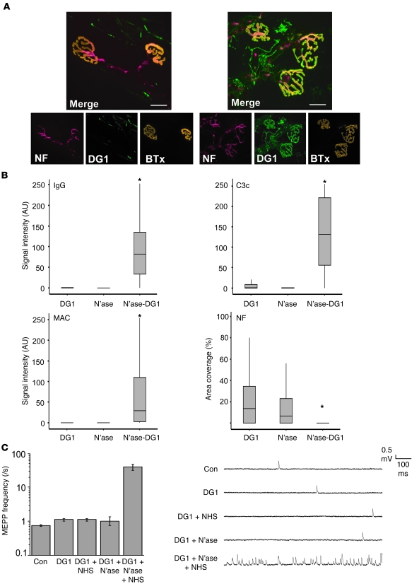
Figure 4. Effect of neuraminidase treatment on anti-GM1 antibody binding and pathogenic activity.
(A) Reconstructed confocal images of DG1 binding at ex vivo GD3s–/– triangularis sterni NMJs. Left: DG1 binding in living tissue is undetectable at the NMJ (stained with αBTx-TRITC). Right: DG1 binding (FITC) following neuraminidase treatment of tissue. DG1 binding overlies the NMJ and is colocalized with the axonal neurofilament staining (Cy5), suggesting that DG1 is binding to the presynaptic aspect (the axon terminal). (B) Ex vivo GD3s–/– hemidiaphragm preparations, as described in Figure 2. Neuraminidase (N’ase) treatment of tissue prior to incubation with DG1 enabled DG1 to bind the NMJ (as shown by IgG deposition), fix complement, and cause a neurofilament loss. *P < 0.05 compared with control tissue incubated in Ringer’s (minus neuraminidase) followed by DG1. (C) Electrophysiology. DG1 and NHS as a source of complement caused a massive increase in MEPP frequency in the ex vivo GD3s–/– NMJ only when applied to neuraminidase-pretreated tissue. Scale bars: 20 μm. Error bars represent SEM for experiments performed in triplicate.

