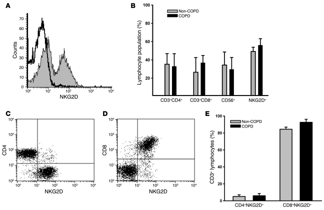
Figure 6. NKG2D receptors are constitutively expressed on pulmonary CTLs.
(A) NKG2D receptor expression on lymphocytes isolated from peripheral lung tissue of a representative COPD patient. Lymphocyte population was defined by forward and side scatter properties. Open histogram represents isotype control staining; filled histogram represents anti-NKG2D staining. (B) Relative abundance of CD3+CD4+, CD3+CD8+, CD56+ (NK), and NKG2D+ lymphocytes in the peripheral lungs of non-COPD and COPD patients. (C and D) CD4+NKG2D+ T cells (C) and CD8+NKG2D+ T cells (D) isolated from peripheral lung tissue of a representative COPD patient gated on CD3+ cells. (E) Relative abundance of CD3+CD4+NKG2D+ and CD3+CD8+NKG2D+ T cells in the peripheral lungs of non-COPD and COPD patients. Values are mean ± SD. n = 5 (non-COPD); 10 (COPD).

