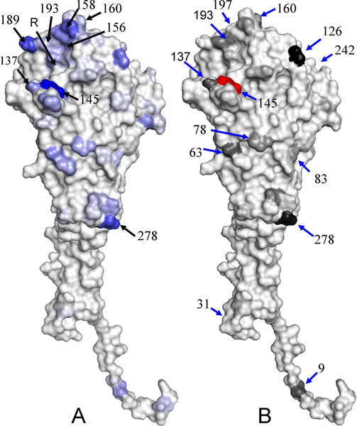
Figure 3.
The distribution of IG values and co-mutation scores on HA structure. (A) The distribution of IG values of 329 amino acids on HA structure (PDB code 1HGF) and the R indicates the receptor binding site. The blue and gray indicate the highest IG value and the lowest IG value, respectively. (B) The structural locations and scores of 12 co-mutation positions of the position 145. These structures are presented by using PyMOL.

