
Figure 1.
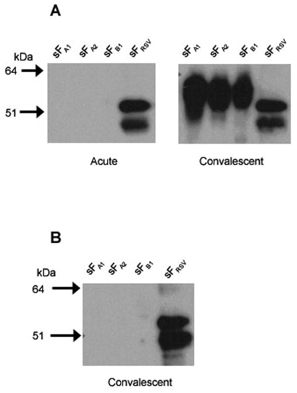
Western blot of presumptive hMPV negative and new infection sera. Purified hMPV sFA1, sFA2, and sFB1 and RSV sF were resolved on 4-12% Nu-PAGE gels and transferred to nitrocellulose membranes. The membranes were probed with human sera as follows: (A) acute and convalescent sera from a patient after the acute sample tested negative but the convalescent sample tested positive for hMPV sF antibody in ELISA, or (B) RSV positive convalescent sera from a patient that was ELISA negative for hMPV sF in both acute and convalescent sera. Bound antibodies were reacted with a peroxidase conjugated goat anti-human IgG and visualized by chemiluminescence.