
Fig 5.
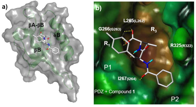
Compound 1 bound to the PDZ domain in an extended conformation as β-peptidomimetics. Compound 1 was docked to the PDZ domain by using Glide (Schrödinger Inc.). The othercompounds adopt similar conformations as that of 1 except 9 and 10. (a) Representations and colors are the same as in Figure 2a. (b) PDZ domain is in surface representation colored by hydrophobicity from brown (hydrophobic) to blue (hydrophilic). Compound 1 is in stick representation. Polar hydrogen atoms are in cyan. hydrogen-bonds between PDZ domain and 1 are in yellow dash lines. The amino acid numbers in bigger fonts are those in Xenopus Dvl PDZ domain and those in smaller fonts correspond to residue numbers in the mouse Dvl1 PDZ domain. The figure was prepared with SYBYL® 7.0 (Tripos, Inc.).