
FIGURE 3. Effect of mutation of Met-6 on the subcellular localization of α2B-AR.
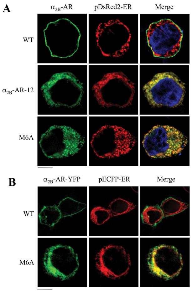
A, co-localization of α2B-AR, α2B-AR-12, and M6A with the ER marker pDsRed2-ER in fixed cells. HEK293T cells were transfected with GFP-tagged α2B-AR, α2b-AR-12, or M6A mutant together with pDsRed2-ER, and the subcellular distribution and co-localization of the receptors with pDsRed2-ER were revealed by fluorescence microscopy as described under “Experimental Procedures.” B, co-localization of α2B-AR and M6A mutant with the ER marker pECFP-ER in live cells. HEK293T cells plated on poly-l-lysine-precoated 35-mm glass-bottom dishes were transfected with YFP-tagged α2B-AR or M6A together with pECFP-ER, and the subcellular localization and co-localization of the receptors with pECFP-ER were obtained in living cells with a Zeiss Axiovert microscope. Green, α2B-AR tagged with GFP (A) or YFP (B); red, the ER markers pDsRed2-ER (A) and pECFP-ER (B); yellow, co-localization of the receptors with the ER; blue, DNA staining by 4,6-diamidino-2-phenylindole (nuclei). The data shown in A and B are representative images of at least three independent experiments. Scale bars, 10 μm.