
FIGURE 4. Effect of mutation of Tyr-12 and Ser-13 on the subcellular localization of α2B-AR.
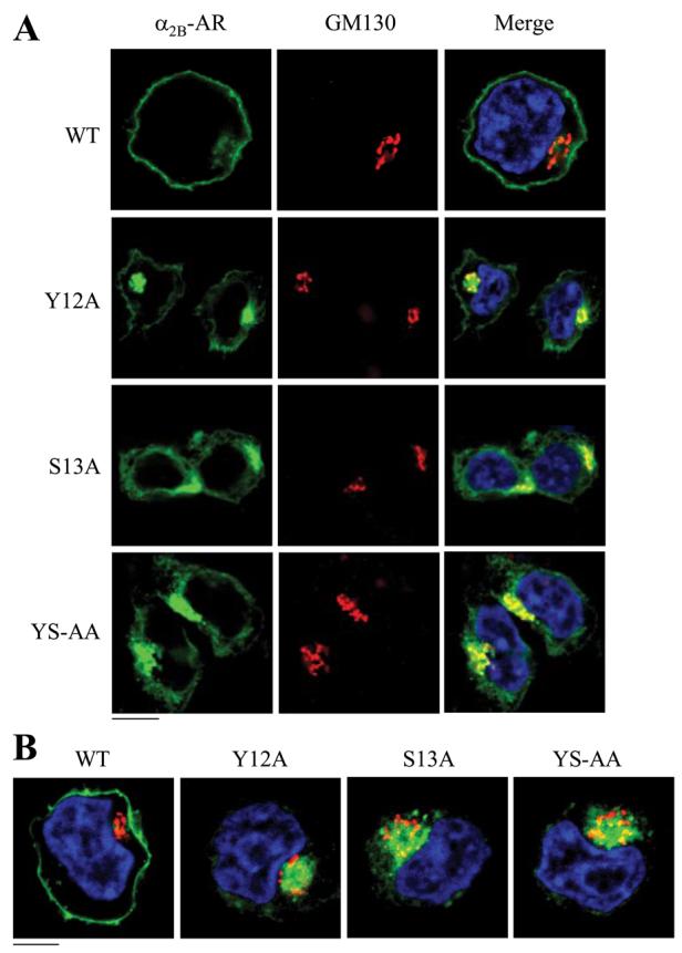
HEK293T cells were transfected with α2B-AR or its mutants Y12A, S13A, and Y12A/S13A and their co-localization with the Golgi (A) and theTGN (B) markers were revealed by fluorescence microscopy after staining with antibodies against GM130 (A) and p230 (B) (1:50 dilution), respectively, as described under “Experimental Procedures.” Green, α2B-AR tagged with GFP; red, the Golgi marker GM130 (A) or the TGN marker p230 (B); yellow, co-localization of the receptors with the Golgi (A) and the TGN markers (B); blue, DNA staining by 4,6-diamidino-2-phenylindole (nuclei). The data shown in A and B are representative images of at least five independent experiments. Scale bars, 10 μm. YS-AA, Y12A/S13A.