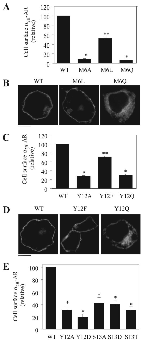
FIGURE 6. Characterization of Met-6, Tyr-12, and Ser-13.
A, effect of mutating Met-6 to hydrophobic Leu (M6L) and non-hydrophobic Gln (M6Q) residues on the cell-surface expression of α2B-AR. B, subcellular localization of α2B-AR and its mutants M6L and M6Q. C, effect of mutating Tyr-12 to Leu (Y12L) and Gln (Y12Q) on the cell-surface expression of α2B-AR. D, subcellular localization of α2B-AR and its mutants Y12L and Y12Q. E, effect of mutating Tyr-12 to Asp (Y12D) and Ser-13 to Asp (S13D) and Thr (S13T) on the cell-surface expression of α2B-AR. HEK293T cells were transfected with α2B-AR or its mutants, their expression at the cell surface was determined by intact cell ligand binding with [3H]RX821002, and subcellular localization was revealed by fluorescence microscopy detecting GFP signal as described in the legend of Fig. 1. The data shown in A, C, and E are percentages of the mean value obtained from cells transfected with WT α2B-AR and are presented as the mean ± S.E. ofthree separate experiments. *,p< 0.05 versus cells transfected with WT α2B-AR; **, p < 0.05 versus cells transfected with M6A (A) and Y12A (C). The data shown in B and D are representative images of three independent experiments. Scalebars, 10 μm.

