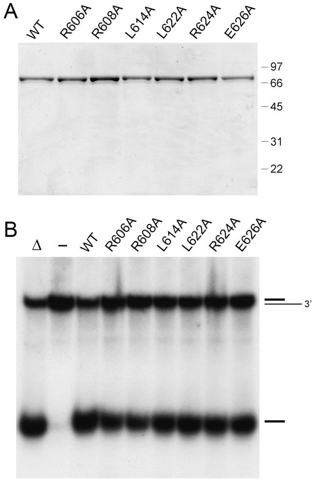
Figure 7.
Alanine scanning of the tetracysteine module. (A) Aliquots (2.5 μg) of recombinant wild-type (WT) UvrD2 and the indicated Ala mutants were analyzed by SDS-PAGE. The Coomassie blue-stained gel is shown. The sizes (kDa) and positions of marker proteins are indicated on the right. (B) Helicase reaction mixtures (10 μL) containing 20 mM Tris-HCl (pH 8.0), 5 mM MgCl2, 50 nM 3′-tailed duplex DNA substrate, 1 mM ATP, and 50 ng of wild-type or mutant UvrD2 as specified were incubated for 5 min at 37 °C. The products were analyzed by native PAGE and visualized by autoradiography. A control reaction without added protein is included in lane -; a control reaction lacking enzyme that was heat denatured prior to PAGE is shown in lane Δ.

