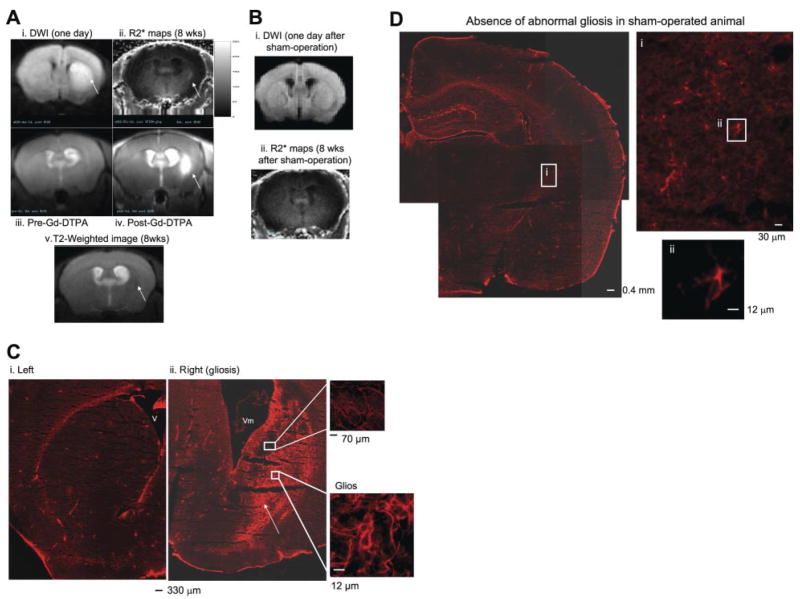
Figure 5.
SPION-gfap detects gliosis in live mice after acute neurological disorder using transcription MRI. We treated C57BL6 mice with BCAO (60 min) to induce BBB leakage. The control group was similarly treated, except there was no vessel occlusion (sham operation). DWI/ADC MRI was obtained at 1 day of reperfusion (Ai, Bi). Eight weeks later, we applied SPION-gfap in an eyedrop solution and acquired T2-weighted MRI from the same mouse (Av), and R2* maps were obtained the next day for the same mouse (Aii) and a sham-operated mouse (Bii). Evidence of BBB leakage is shown by pre- and post Gd-DTPA (0.1 mmol/kg, i.v.) and T1-weighted MRI in vivo (Aiii, iv) 9 wk after BCAO. In the postmortem samples obtained after all in vivo data acquisitions in the same mouse, a cohort of GFAP-expressing cells (Cy-3-labeled anti-rabbit IgG and rabbit anti-GFAP) is located in the right (Cii), but not in the left striatum of postmortem samples from the same mouse (Ci). A normal pattern of GFAP-expressing cells in a sham-operated mouse (n=4) is shown in D. Arrows indicate DWI hyperintensity (Ai), possibly injured tissue (Av), elevated R2* signal (Aii), BBB leakage (Aiv), and gliosis (Cii). Color scale = 0–250 s−1 (A).

