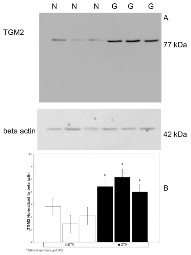
Figure 1.
Chemiluminescent detection of TGM2 protein in normal and glaucomatous human TM cells. (A) Total protein was collected from three normal (N) and three glaucomatous (G) cell lines and electrophoresed in SDS-PAGE gels followed by Western immunoblot analysis for TGM2 (77 kDa). All cell lines express TGM2 proteins. Protein levels of TGM2 were higher in GTM than in the NTM cell lines. β-Actin was used as an internal loading control. (B) Densitometric readings of TGM2 normalized to β-actin for three NTM and three GTM cell lines. *Statistical difference at the P < 0.005 level (±SD).

