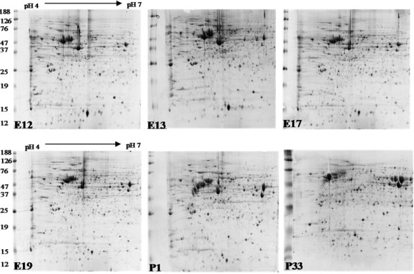
Figure 1.

Representative 2D protein profiles from embryonic and post hatch chick retina. Retinal protein was extracted in 40 mM ammonium bicarbonate and 1 mg of extracted protein was separated in the first dimension between pH 4 to 7 on an 18 cm IPG strip, and, in the second dimension on a 12% polyacrylamide gel (25 cm × 20 cm). Proteins were visualised with Coomassie blue.
