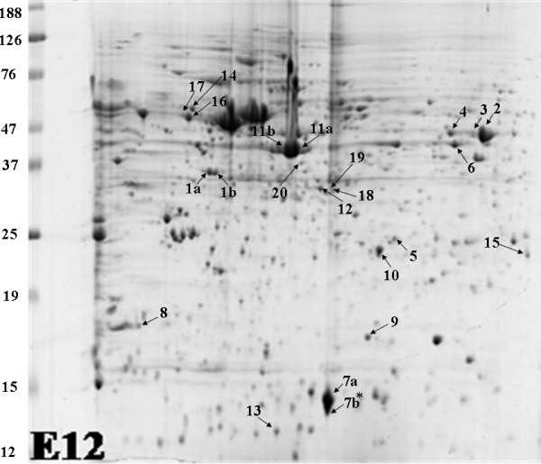
Figure 3.

Chick retinal proteins were subjected to 2D gel electrophoresis, separated through pH 4–7 and stained with Colloidal Coomassie blue. A representative 2-D gel image with the spots identified by MS analysis is shown. * This spot resolved as two distinct spots at older ages, two spots were identified by MS as FABP7.
