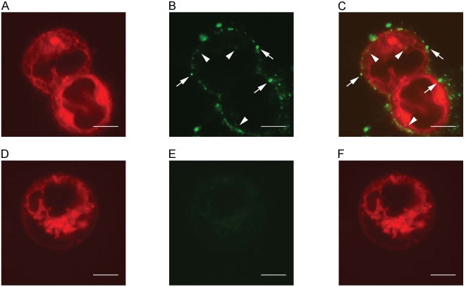
Figure 6. ESMVs transfer GFP.
ESCs without the GFP transgene were labeled with DiD and then incubated with ESMVs containing GFP. All confocal images were taken using a 100×, 1.4 NA objective with the pinhole set to 1 airy unit. (A) DiD signal from ESCs incubated with ESMVs. (B) GFP signal from ESCs incubated with ESMVs. Arrows indicate punctate signal, likely representing docked vesicles. Arrowheads indicate diffuse signal, likely from the diffusion of GFP inside the cell or from the production of newly translated GFP. (C) Overlay of A+B. (D) DiD signal from control ESCs without ESMVs. (E) No GFP signal can be detected in the absence of ESMVs. (F) Overlay of D+E. All scale bars are 5 μm.

