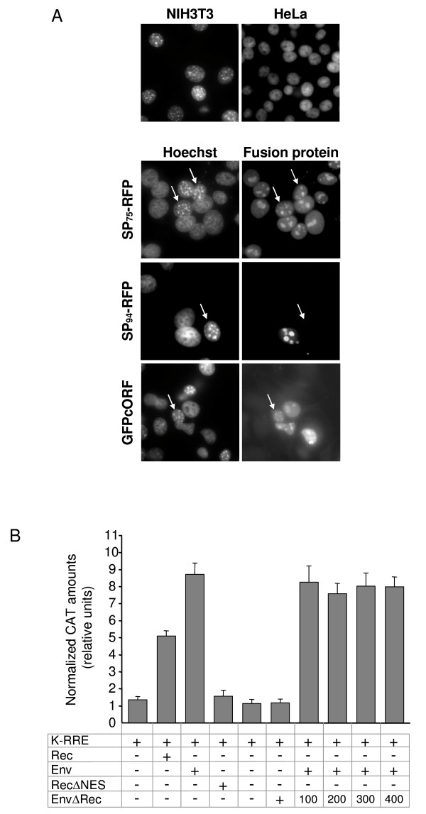
Figure 8.
Functional differences between HML-2 SP and Rec. (A) Heterokaryon assay. HeLa cells were transfected with SP75-RFP, SP94-RFP or GFPcORF expression plasmids and co-cultured with mouse NIH3T3 cells. Cells were subsequently treated with 100 μg/ml CHX for 1 hour. After induction of cell fusion, cells were fixed and immunostained. Counterstaining with Hoechst 33258 served to distinguish human from mouse nuclei (left panels). Arrows indicate NIH3T3 nuclei that accumulated SP94/75-RFP or GFPcORF after syncytia formation (right panels). (B) Measurement of HML-2 SP RNA export activity by quantitative determination of chloramphenicol acetyltransferase (CAT). HeLa cells were co-transfected with the CAT reporter plasmid pDM128/K-RRE (K-RRE) together with either Rec, Env, RecΔNES, or EnvΔRec expression plasmids. Histogram bars represent normalized amounts of CAT as determined by CAT ELISA, indicating export of unspliced CAT mRNA to the cytoplasm. Where indicated, 400 ng of Env expression plasmid were co-transfected with increasing amounts of EnvΔRec plasmid (100, 200, 300 or 400 ng), and with constant amounts of the CAT reporter vector. All values are given as the mean of at least 4 independent experiments. Error bars indicate the SEM.

