FIGURE 1.
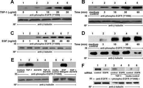
TSP1 activates EGFR in A431 cells. A431 cells were exposed for 0.5 h to increasing concentrations of TSP1 or media alone (A) or exposed for increasing times to a fixed concentration of TSP1 (30 μg/ml or 214 nm) or media alone (B). In other experiments, A431 cells were exposed for 10 min to increasing concentrations of EGF or media alone (C) or exposed for increasing times to a fixed concentration of EGF (100 ng/ml or 16.7 nm) or media alone (D). E, cells were incubated for 0.5 h with TSP1 (30 μg/ml i.e. 214 nm), for 10 min with EGF (100 ng/ml i.e. 16.7 nm), or media alone, in the presence or absence of the EGFR-selective tyrphostin, AG1478 (5 μm). F, cells were transfected with control or EGFR-targeting siRNAs, and after 48 h were lysed and the lysates processed for EGFR immunoblotting (lanes 1 and 2). After transfection with either control or EGFR-targeting siRNAs, cells were incubated with TSP1 (lanes 3 and 4) or media alone (lanes 5 and 6). Cells were lysed and the lysates processed for immunoblotting with anti-phospho-EGFR (Tyr-1068) antibodies. To indicate protein loading and transfer, blots were stripped and reprobed with anti-β-tubulin antibody. IB, immunoblot; IB*, immunoblot after stripping. Each of these blots is representative of >2 experiments.

