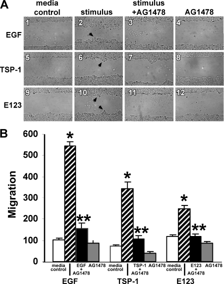
FIGURE 6.
TSP1 increases cell migration through EGFR activation. A431 cells were cultured to confluence in the wells of 24-well plates, after which they were wounded with a pipette tip, washed to remove cellular debris, and incubated for 48 h with EGF (10 ng/ml or 1.67 nm), TSP1 (30 μg/ml or 214 nm), E123 (3.8 μg/ml or 214 nm), or media alone in the presence or absence of the EGFR selective tyrphostin, AG1478 (5 μm)(n = 6). At 48 h, cellular migration into the wound was photographed in triplicate and quantified. A, representative photographs of wounded monolayers after 48 h of incubation with EGF, TSP1, E123, or media alone in the presence or absence of AG1478. Arrows in panels 2, 6, and 10 indicate increased cell migration into the wound. Magnification, ×40. B, vertical bars represent mean (±S.E.) migration into the wound at 48 h after incubation with EGF, TSP1, E123, or media alone in the presence or absence of AG1478. For each condition, n = 6. *, significantly increased compared with the media control at p < 0.05. **, significantly decreased compared with the stimulus alone at p < 0.05.

