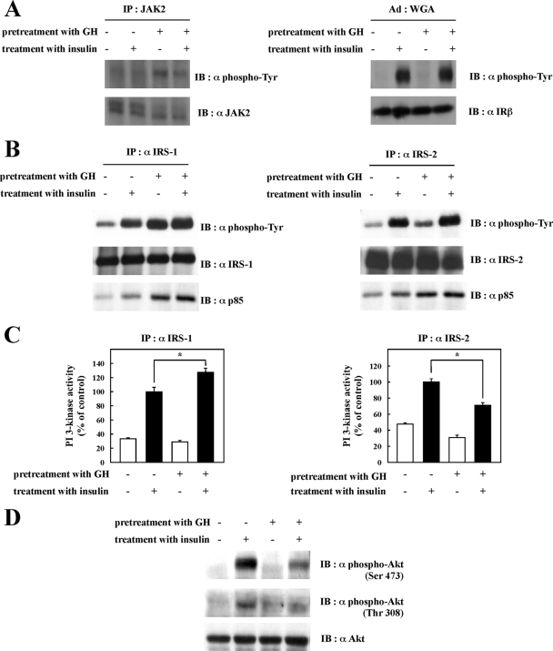
FIGURE 3.
Effects of chronic GH pretreatment on insulin signal activation. After being serum-starved for 2 h, 3T3-L1 adipocytes were pretreated with or without 100 nm GH for 24 h and then treated with or without 0.1 nm insulin for 5 min. Cells were solubilized with Tris/Triton lysis buffer. A, whole cell lysates were immunoprecipitated (IP) with anti-JAK2 antibody, and the immunoprecipitates were subjected to immunoblotting (IB) with anti-phosphotyrosine or anti-JAK2 antibody (left panel). Insulin receptor was semi-purified with wheat germ agglutinin (WGA)-agarose from whole cell lysates. Semi-purified insulin receptor was separated by SDS-PAGE and immunoblotted with anti-phosphotyrosine antibody or anti-insulin receptor-β antibody (right panel). B, whole cell lysates were immunoprecipitated with anti-IRS-1 antibody or anti-IRS-2 antibody. Immunoprecipitates were separated by SDS-PAGE and immunoblotted with anti-phosphotyrosine antibody, anti-IRS-1 antibody, anti-IRS-2 antibody, or anti-p85 PI 3-kinase antibody. C, whole cell lysates were immunoprecipitated with anti-IRS-1 antibody or anti-IRS-2 antibody. PI 3-kinase activity in the immunocomplexes was measured as described under “Experimental Procedures.” PI 3-kinase activities were quantified, and the results are presented as the means ± S.E. of three independent experiments. The difference between insulin-stimulated cells with and without GH pretreatment is significant with p < 0.05 (*). D, whole cell lysates were separated by SDS-PAGE and immunoblotted with anti-phospho-Akt (Ser-473) antibody, anti-phospho-Akt (Thr-308) antibody, or anti-Akt antibody.

