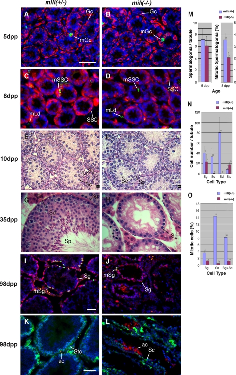
FIGURE 1.
The defect of mili mutant in the self-renewal of spermatogonial stem cells. A-D, testicular sections of 5-day-old (A and B) and 8-day-old (C and D) mili+/- (A and C) and mili-/- (B and D) mice stained with EE2 antibody to outline spermatogonia (red), anti-phosphohistone 3 antibody to label mitotic cells (green), and DAPI to label DNA (blue). The mili+/- and mili-/- testes contain the same number of spermatogonia, as quantified in M, but the mitotic rate in the mili-/- (2.1 ± 0.4%) is noticeably lower than that of mili+/- spermatogonia (3.6 ± 0.4%). Note that all spermatogonia (Sg) at 5 dpp are germ line stem cells. Gc, gonocyte; mGc, mitotic gonocyte; SSC, spermatogonial stem cell; mSSc, mitotic SSC; mLd, mitotic Leydig cells. E-H, hematoxylin/eosin-stained testicular sections of 10-day (E and F) and 35-day (G and H) old mili+/- (E and G) and mili-/- (F and H) mice. Sc, spermatocyte; Sd, spermatid; Sp, sperm. In mili-/- testes, most germ cells are arrested as spermatogonia, with a tiny fraction of germ cells differentiating into spermatocytes. However, no spermatid is detected. This phenotype persists through up to 180 days and was qualified at 98 days in N. I and J, testicular sections of 98-day-old mili+/- (I) and mili-/- (J) mice stained for EE2 (red), phosphohistone 3 (green), and DNA (blue). The mitotic rate of mili-/- spermatogonia is significantly lower than that of the mili+/- spermatogonia, as quantified in O. mSg, mitotic spermatogonia. K and L, testicular sections of 98-day old mili+/- (K) and mili-/- (L) mice stained with BC7 antibody to mark spermatocytes (green) and terminal deoxynucleotidyltransferase-mediated dUTP nick end labeling to mark apoptotic cells (ac; red) and DNA (blue). Stc, Sertoli cell. Note that drastic apoptosis occurs in the arrested mili-/- germ cells. Bars in A, I, and K denote 50 μm for A-H, I and J, and K and L, respectively. M-O, quantitative comparisons between mili+/- and mili-/- testes with regard to the number of spermatogonia per tubule at 5 dpp (M) and 98 dpp (N), and mitotic index of spermatogenic cells at 8 dpp (M) and 98 dpp (O).

