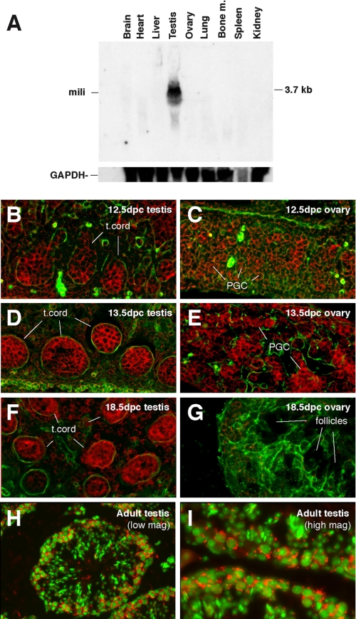
FIGURE 2.
The expression of mili mRNA during mouse germ line development. A, a Northern blot containing total RNAs prepared from various adult tissues as indicated and probed for the mili mRNA. B-I, in situ mili RNA hybridization of embryonic and adult testes and ovaries at specific stages as indicated. Red, mili mRNA; green in B and G, laminin, which outlines testicular cord (t. cord) in B, G, and F; green in H and I, DNA (stained with DAPI).

