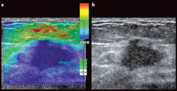
Figure 5. Imaging elastography of a breast tumour.
Tissue imaging elastography is a spatial ‘visual’ qualitative measurement of the stiffness of a tissue that is generated by extrapolating tissue viscoelastic characteristics from ultrasound wave reflection in real-time. Photographs of sonoelastography images compare an elastogram image (a) with a B mode ultrasound scan (b) of a breast tumour170. Ultrasound imaging elastography, as shown here, is an in situ mechanical imaging method that could improve the sensitivity and the specificity of breast cancer detection and may be a useful tool to advance our understanding of the link between mammographic density and the matrix materials properties of the breast. Image courtesy of A. Thomas & T. Fischer, Charité, Berlin, Germany.

