
FIGURE 4. PSD-95 reduces D1 surface expression and promotes intracellular D1 localization.
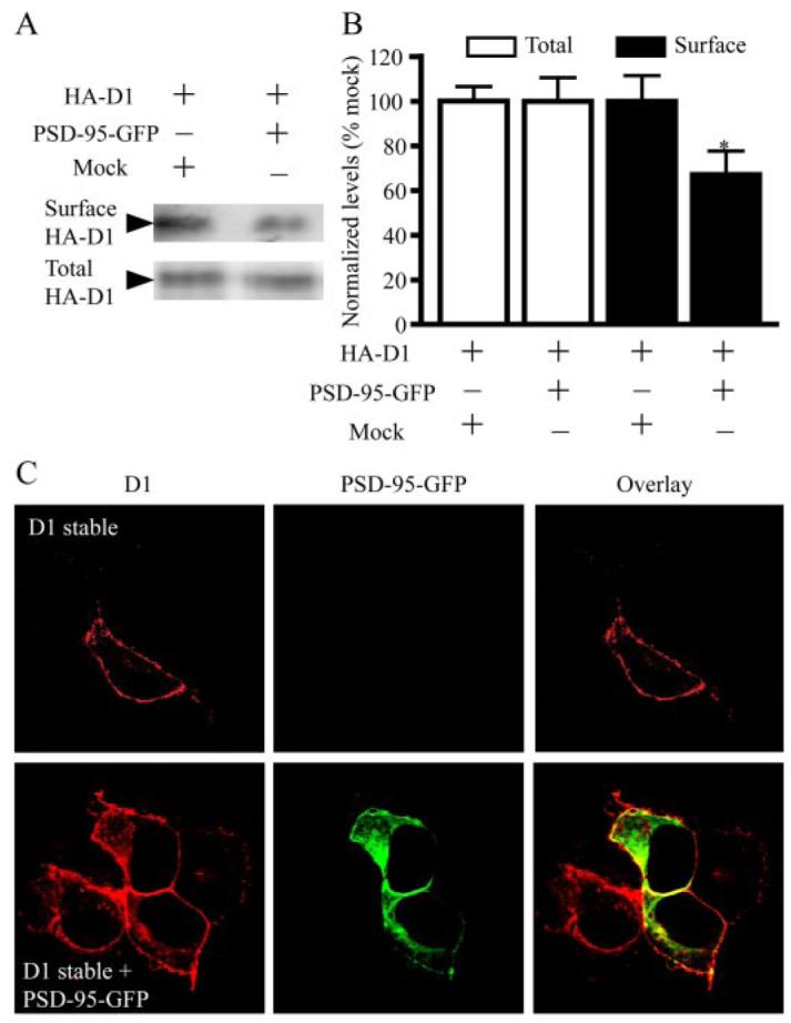
A, reduced surface D1 expression by PSD-95. Cell surface biotinylation experiments were performed on HEK293T cells transiently trancfected with HA-D1 and an empty vector or HA-D1 and PSD-95-GFP. Biotin-labeled surface D1 and total D1 were analyzed by Western blots. B, quantification of surface and total D1 levels. Mean ± S.E. represents three independent experiments. *, p < 0.05, Student’s t test. C, increased presence of intracellular D1 in PSD-95-GFP co-expressing cells. D1 stable HEK293 cells were transiently transfected with a mock vector or PSD-95-GFP. Following immunostaining, fluorescence was visualized by confocal microscopy. For this and the following figures, confocal images shown were single focal plane scannings through the middle of cells.