Fig. 3.
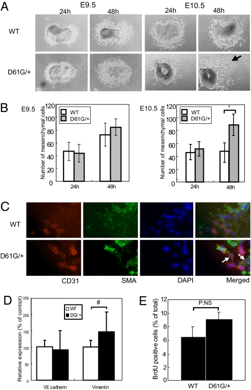
Sustained EMT in D61G/+ EC cushions. (A, B) Explant assays using E9.5 (Left) or E10.5 (Right) EC from WT and DG/+ embryos. Mesenchymal cells were counted at 24 and 48 h. Note that EC from E10.5 DG/+ embryos produce more mesenchymal cells (arrow). Values represent mean ± SD. n = 8. *, P < 0.01, by 2-tailed student's t test. (C) Excess mesenchymal cells are caused by sustained EMT. Cushion explants at E10.5 were stained with the indicated antibodies and DAPI. CD31 and αSMA double-positive “transitional” cells (white arrows) are found only in DG/+ explants. (D) Quantitative RT-PCR of VE-cadherin and vimentin. Values represent mean ± SD. n = 8. #, P < 0.05, by 2-tailed student's t test. (E) Proliferation of cushion mesenchymal cells. Explants (7/genotype) exposed to BrdU for 6 h were immunostained with BrdU antibodies, and BrdU+ cells were counted. NS, not statistically significant.

