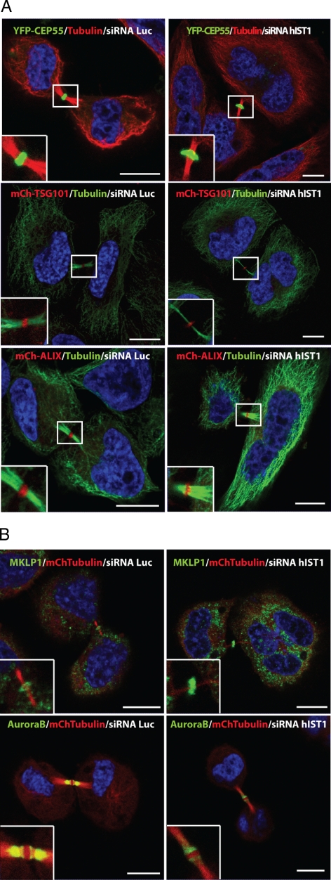
Figure 9.
Characterization of the midbody composition in cells that lack hIST1. (A) Representative micrographs describing midbody localization of YFP-CEP55 (green), mCh-TSG101 (red), or mCh-ALIX (red) in cells stably expressing these fusion proteins and treated with siRNA targeting luciferase or hIST1. After siRNA treatment, cells were fixed and stained with α-tubulin and Alexa594 (red) or Alexa488 (green) conjugated secondary antibodies. (B) Similarly, HeLa cells stably expressing mCh-Tubulin (red) treated with siRNA targeting luciferase or hIST1 were immunostained for MKLP1 or AuroraB (green). In all cases, enlargements depict the midbody localization of the studied proteins. DNA is shown in blue. Bar, 10 μm.

