Figure 7.
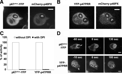
Accumulation of NADPH oxidase probes on the phagosome during IgG-Zym phagocytosis in PLB-985 neutrophils treated with wortmannin or DPI. Translocation of p67phox-YFP (A) and YFP-p47PRR (B) during IgG-Zym phagocytosis in transgenic PLB-985 neutrophils coexpressing mCherry-p40PX in the presence of 100 nM wortmannin. Letters m and n indicate individual internalized phagosomes. PLB-985 granulocytes expressing p67phox-YFP or YFP-p47PRR were pretreated with 10 μM DPI at 37°C for 15 min, followed with synchronized phagocytosis as described in Materials and Methods. NADPH oxidase activity was measured in the presence of luminal, SOD, and catalase. Results are expressed as total relative light unit (RLU) values more than 45 min, measured at 1-min intervals. (C) Values represent the mean ± SD of triplicate determinations. Live image was monitored using confocal microscopy as described in Figure 3A. (D) Arrows indicate presealing (newly forming phagosomes) and letters o and p indicates internalized phagosome. Time-lapse confocal microscopy was used as described in Figure 3A. Bar, 5 μm.

