Figure 5.
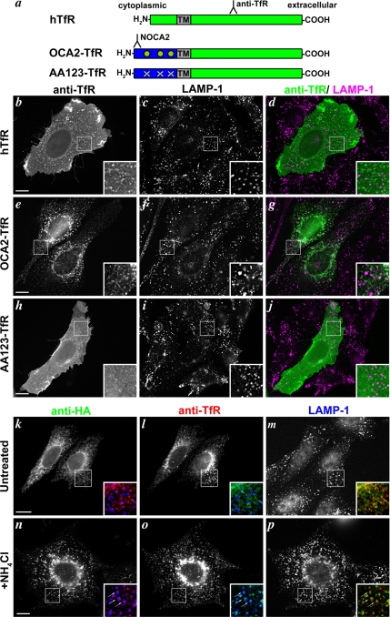
OCA2 dileucine motifs are sufficient for lysosomal localization in nonmelanocytes. (a) Schematic of OCA2-transferrin receptor chimeras. Green, hTfR sequences; blue, cytoplasmic N terminus of human OCA2; TM, hTfR transmembrane domain; green circles, intact OCA2 dileucine motifs; white X's, disrupted dileucine motifs. Binding regions of relevant antibodies are indicated. The NOCA2 epitope does not overlap the dileucine motifs. (b–j) Chinese hamster ovary cells were transfected with hTfR (b–d), OCA2-TfR (e–g), or AA123-TfR (h–j), and the transgenic proteins were localized at steady-state by anti-hTfR staining (b, e, and h). The lysosomal marker LAMP-1 is stained in c, f, and i, and images are merged in d, g, and j with anti-hTfR in green and anti-LAMP-1 in magenta. Insets, 4X magnification of boxed regions. (k–p) Chinese hamster ovary cells were cotransfected with wild-type OCA2-HA (k and n) and OCA2-TfR (l and o) and incubated with (n–p) or without (k–m) 50 mM NH4Cl before fixation in order to inhibit lysosomal degradation. Lysosomes are marked by LAMP-1 staining (m and p). Insets show 3X magnifications of two-color merged images of OCA2-HA (green), OCA2-TfR (red), and LAMP-1 (blue). Arrows point to regions of overlap among the three proteins (n–p). Bar, 10 μm.

