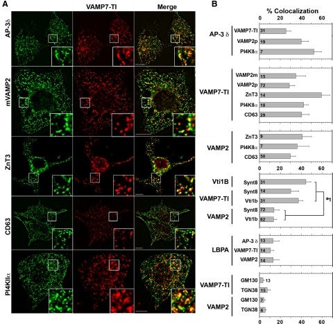
Figure 1.
AP-3 synaptic vesicle and lysosomal cargoes selectively colocalize in PC12 Cells. (A) Fixed PC12 cells immunostained for VAMP7-TI and indicated protein. Cells were imaged by wide-field deconvolution microscopy. Bar, 5 μm. (B) Quantification of colocalization between AP-3 and synaptic vesicle (VAMP2) or lysosomal cargo (VAMP7-TI and PI4KIIα); VAMP7-TI and synaptic vesicle (VAMP2 and ZnT3) or lysosomal cargo (PI4KIIα and CD63); VAMP2 and synaptic vesicle or lysosomal cargo. Cognate SNARE interactions and late endosomal marker LBPA illustrate the minimal contribution of late endosomes to the observed synaptic vesicle/lysosomal cargo colocalization. Golgi markers GM130 and TGN38 were used as negative controls to assess background levels of colocalization. Number of analyzed cells is indicated at the bottom of each bar graph. Asterisk represents p values for comparison of colocalization between either Vti1b or VAMP7-TI and either Syntaxin 8 or Vti1b, with colocalization between VAMP2 and Syntaxin 8 or Vti1b. All p < 0.0001. One-way analysis of variance (ANOVA) Student–Newman–Keuls multiple comparison. Representative images of these quantifications are presented in Supplemental Figures 2–4.

