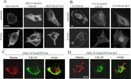
Figure 4.
Force-induced redistribution of FilGAP. Wild-type and FLNa-knock down HEK (A) and wild-type and FLNa-knockdown NIH 3T3 cells (B) were plated on fibronectin-coated glass coverslips, transfected with HA-tagged FilGAP or HA-tagged FilGAPΔCC for 48 h, and then either untreated or subjected to force. FilGAP was localized by staining the cells with anti-HA antibody. (C) FLNa-knockdown HEK cells were double-transfected with HA-tagged FilGAP and DsRed-FLNa construct, incubated with collagen beads, and subjected to force. (D) Double-transfection of FLNa-knockdown HEK cells with HA-tagged FilGAP and DsRed-FLNaΔ23 constructs. Transfected cells were incubated with collagen beads and subjected to force. Cellular distribution of DsRed-FLNa was directly visualized (red), whereas FilGAP was detected with anti-HA antibody (green). Bar, 10 μm.

