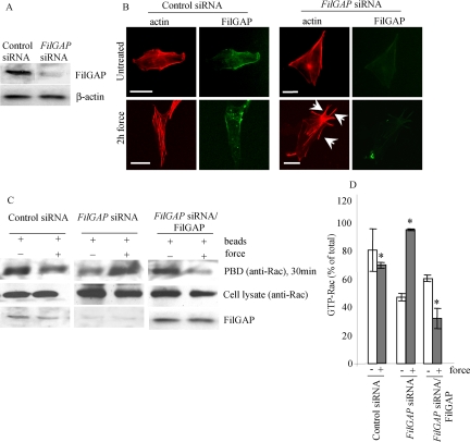
Figure 6.
FilGAP suppresses force-induced lamellae. (A) Immunoblot showing that FilGAP is depleted 48 h after siRNA treatment of NIH 3T3 cells. (B) 3T3 cells were plated on fibronectin-coated glass coverslips and treated with siRNA targeting endogenous FilGAP as described in A. Cells were loaded with equal amount of collagen-coated beads and either left untreated or subjected to force. FilGAP and F-actin were localized by staining the cells with anti-FilGAP antibody (green) and rhodamine-phalloidin (red), respectively. Lamellipodia are indicated by arrows. Bar, 20 μm. (C) 3T3 cells were treated with siRNA alone as described in A or, after 48 h, they were transfected with full-length FilGAP. Cells were then subjected to force as described in B. Cell extracts were incubated with GST-PBD and immobilized on glutathione-Sepharose beads. The amount of Rac in cell lysates before pull-down; GST-PBD–bound Rac was detected by immunoblot. (D) Intensities of GST-PBD–bound Rac bands were measured using ImageJ densitometry software. Numbers represent ratios relative to the levels of Rac in cell lysates before pull-down and are expressed as the means and SDs of three independent experiments. *p < 0.05 versus respective untreated control.

