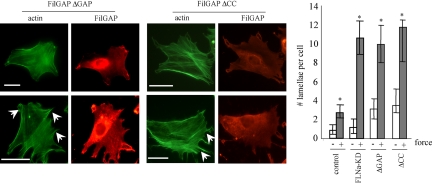
Figure 7.
Requirement of FilGAP activity and binding to FLNa for suppression of force-induced lamellae. 3T3 cells were either left untransfected or transfected with HA-tagged FilGAPΔGAP (dominant negative) or FilGAPΔCC, lacking coiled-coil domain. After 48 h, cells were incubated with collagen-coated beads and subjected to mechanical force or left untreated. FilGap and F-actin were visualized by staining the cells with anti-HA antibody (red) and FITC-phalloidin (green), respectively; lamellipodia are indicated with arrowheads. Bar, 20 μm. Numbers of lamellipodia were counted. The figures show means and SDs of three microscopic fields, with an average of 50 cells per each field. *p < 0.05 versus respective untreated control.

