Figure 9.
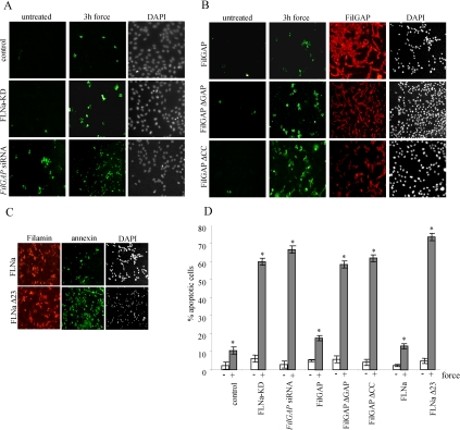
Inhibition of force-induced apoptosis. (A) Control HEK, FLN-KD HEK, and HEK cells pretreated with FilGAP siRNA for 48 h were incubated with collagen-coated beads and subjected to mechanical force or left untreated. Apoptotic and necrotic cells were detected with annexin-V-Fluos (green) and PI (data not shown), respectively, and the total amount of cells in each microscopic field was visualized by DAPI staining. (B) HEK cells were plated on fibronectin-coated glass coverslips and pretreated with FilGAP siRNA. Forty-eight hours later, cells were transfected with HA-tagged FilGAP, FilGAPΔGAP, and FilGAPΔCC constructs. After 24 h, cells were subjected to mechanical force or left untreated, and apoptotic cells were visualized as described in A. Transfection efficiency was determined by staining the cells with anti-HA antibody (red). (C) HEK FLNa-KD cells were transfected with either full-length DsRed-FLNa or DsRed-FLNaΔ23 construct, subjected to force and stained as described in A. (D) Quantification of apoptosis. For all cells apoptosis was measured as the percentage of annexin-V-Fluos–positive cells out of total number of cells in microscopic field, multiplied by transfection efficiency. Data are means and SDs of three microscopic fields, an average of 80 cells per field. *p < 0.0002 versus respective untreated control.

